Back to: O level Biology NOTES Uganda syllabus

Parts and functions of the urinary system
1) Aorta
It carries oxygenated blood with all food nutrients to the kidney.
2) Renal artery:
This arises from dorsal aorta. It brings blood containing excretory products to the kidney.
3) Renal vein:
It carries filtered blood from the kidney to the posterior vena cava.
4) Ureter:
These are two narrow tubes arising from hilum of each kidney. They connect the kidneys to the urinary bladder. They transport urine to the urinary bladder.
5) Urinary bladder:
It is a thick walled elastic sac-like structure which stores urine.
6) Sphincter muscle:
These muscles are elastic thus can contract and relax to control urine flow.
7) Urethra:
It is a passage for urine to the outside of the body.
THE KIDNEY

The kidneys are solid bean-shaped structures and they occur in pairs in mammals. They are reddish-brown in colour enclosed in a transparent membrane and attached to the back of the abdominal cavity.
The kidney tissue consists of many capillaries and renal tubules connected together by connective tissue. The kidney has two major parts.
- The cortex which is a dark outer part. It consists of the Bowman’s capsule which is responsible for ultra-filtration of blood passing across it.
- The medulla, which is a lighter inner, part. It is made up of many cone-shaped portions called pyramids.
The pelvis is the area where the ureter leaves the kidney.
The kidney performs three major functions in the body.
- It carries out excretion.
- It carries out the function of osmoregulation.
- It contains endocrine glands, which secrete hormones.
The kidney is made up of several microscopic structures (functional units) called nephrons where the actual excretion and osmoregulation takes place.
THE NEPHRONE
This is the functional unit of the kidney. It carries out the function of excretion and osmoregulation in the kidney.
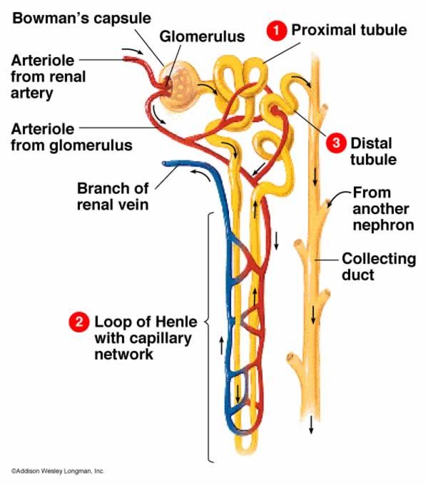
The nephron consists of a cup-shaped structure known as the bowmans’ capsule. Blood comes to the nephrone through the afferent vessel, which is a branch of the renal artery, and it leaves through the efferent vessel.
The efferent vessel joins many other efferent vessels from other nephrones to form the renal vein.
In the bowmans’ capsule the afferent vessel divides to form capillaries.
The capillaries are highly coiled and they form a knot called glomerulus.
Leading from the bowman’s’ capsule is a highly coiled tube known as proximal convoluted tubule. This is continuous with a U shaped tubule called loop on Henle.
The loop is divided into the descending loop and ascending loop.
From the loop of Henle the tube becomes highly coiled to form the distal convoluted tubule which leads to the collecting duct.
Structure of the nephron

Parts of nephron
- Bowman’s capsule:
It contains a dense-network of capillaries called glomerulus. The glomerulus is formed from the wider arteriole of renal artery called afferent arteriole. It is located in the cortex.
The Bowman’s capsule serves the function of filtering small molecules in blood such as urea glucose, etc. through a process called ultra-filtration.
Adaptations of the glomerulus to ultra-filtration
i) Having high blood pressure that forces small molecules out of the glomerulus. This is due to the afferent arteriole being wider than the efferent.
ii) Having many capillaries that give it a large surface area for ultra-filtration.
iii) Having a semi permeable membrane that can allow any small molecule to pass through.
Adaptations of the Bowman’s capsule to collect the filtrate
i) Possession of cup-shaped structure which enables it to collect the filtrate.
ii) Having a porous upper membrane that easily allows filtration.
iii) Having a large volume that can accommodate more filtrate.
- Proximal convoluted tubule:
This is a site where re-absorption of useful materials such as glucose and some small amino acids and water from glomerular filtrate back to blood takes place. - Loop of Henle:
It’s made up of a descending (going down) limb and an ascending (going up) limb. The main function of the loop of Henle is to make the tissue fluid in the medulla more concentrated than the glomerular filtrate in the nephron so that water needed in the body is reabsorbed. It’s known to cause the retention of water. This is one way of conserving water in camel because of its extremely long loop of Henle. - Distal convoluted tubule:
It chiefly re-absorbs salts like chloride ions together with water, leaving a concentrated liquid now called urine which passes down to collecting ducts. - Collecting duct:
This duct carries urine from the distal tubule to the pelvis of kidney. It allows outward movement of water thus conserving it.
Adaptations of the nephron to re absorption
i) Having a thin membrane (one cell thick) for easy diffusion of materials.
ii) Having micro villi to increase the surface area for re absorption.
iii) Having numerous mitochondria to provide energy for active reabsorption.
URINE FORMATION
The process of urine formation takes place in the nephrone. It occurs in two phases.
- Ultra-filtration.
- Selective re-absorption.
Ultra filtration
Much blood comes from the afferent vessel into the glomerulus than that which leaves through efferent because the afferent vessel is larger than the efferent vessel.
This generates pressure in the blood capillaries of the glomerulus forcing small molecules to filter out of the blood capillaries to form the glomerular filtrate.
Blood in the renal artery contains proteins, red blood cells, white blood cells, urea, water, salts, amino acids and vitamins.
In the glomerulus, small molecules filter out by ultra filtration to form the glomerular filtrate. This filtrate contains glucose, urea, water, salts and vitamins.
Proteins and blood cells do not filter out because they have bigger molecules, which cannot pass through the walls of the glomerulus.
The filtrate formed moves from the Bowman’s capsule through the capsular space to proximal convoluted tubule where selective reabsorption starts to occur.
Diagrammatic illustration of ultrafiltration

Selective reabsorption
- In the proximal convoluted tubule:
- Most of the food materials are re absorbed into the blood capillaries by active transport e.g. all the glucose, vitamins, some salts like sodium chloride and even some water is re absorbed by diffusion.
- In the loop of Henle:
- As the filtrate flows down the descending limb, water is re absorbed back into the capillaries by osmosis leading to increased concentration of the filtrate down the descending limb.
- As the filtrate ascends, the thick ascending limb of loop of Henle, salts like Na and K are reabsorbed by active transport. This leads to a decrease in concentration of the glomerular filtrate in the ascending limb.
- In the distal convoluted tubule:
- Selective re absorption of salts by diffusion occurs.
- In the collecting duct:
- Water is lost to the highly concentrated medulla tissues by osmosis from which later the remaining filtrate is urine which goes via the ureter and temporarily stored in the urinary bladder.


- There are proteins in blood and there is none in urine because proteins are not filtered out of the blood vessels into the glomerulus due to the large size of their molecules.
- Urea is more in urine than in blood because it is filtered out of blood and it is not reabsorbed back in the blood.
- Water is more in urine than in blood because it is used to dissolve urea.
- However the relative amounts of water in urine and in blood varies depending on the amount of water in the body, amount of solutes in the body, temperature and body activity.
- There is glucose in blood and no glucose in urine because glucose is reabsorbed from the glomerular filtrate back into the blood.
- Salts like chlorides and sodium ions are more in urine than in blood. This is because they are in excess and they are not reabsorbed back into the blood. Because of this they tend to concentrate in urine.
