Back to: O level Biology NOTES Uganda syllabus
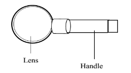
1.HAND LENS:
A normal hand lens is a convex lens mounted in a frame. It is placed a shorter distance of about 5cm from the eye and the object.
Determination of magnification using a hand lens
Magnification refers to how much larger the object appears compared to its real size.

Example
Calculate the magnification of an object, which is 10cm tall whose image appears to be 20cm tall.
Solution
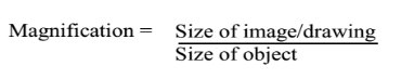
Magnification = 20/10
=x2
2.MICROSCOPES
There are two types of microscopes i.e.
- The electron microscope which uses a beam of electrons.
- The compound light microscope
THE COMPOUND LIGHT MICROSCOPE
It is called so because it uses a beam of light to view objects and has more than one convex lens. it is used in hospitals, schools and some industries.
Structure of a compound light microscope.
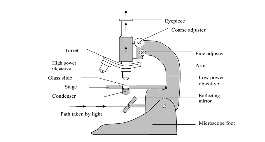
FUNCTIONS OF THE DIFFERENT PARTS1.
1.Eye Piece:
Enables one to view the specimen
It magnifies the image from the objective lens.
2.Barrel:
Provides support for the eye piece and objective lens.
3.Nose piece/ turret/:
It holds the objective lenses in position
Can be rotated to position a particular lens required for a particular magnification.
4.Stage:
It is where a prepared slide is placed for observation.
5.Mirror:
It reflects light from external source through the specimen.
6.Stand / Base:
Supports instrument in on a flat surface.
7.Diaphragm:
Regulates the amount of light passing through the specimen.
8.Condenser:
Concentrates the light reflected by the mirror through the object / specimen on the stage.
9.Arm:
Used for carrying the instrument.
10.Clip:
Keeps the slide firmly on the stage.
11.Coarse adjustment knob:
Used for focusing of the object under study.
12.Fine adjustment knob:
Brings specimen into a sharp clearer focus (final focusing).
13.Objective lens:
Magnifies the specimen under study.
They are normally two or three. Low power (shortest), medium power and high power (longest).
Care of a microscope
The microscope is very delicate, expensive instrument which is very useful in the study of biology. Thus it should be handled carefully doing the following;
- It should be carried with both hands.Should never be dropped.
- Always kept in an upright position
- Only wipe the lens with soft lens tissue.
- It should always be kept in its special designed box.
Determination of magnification of a microscope.
Magnification refers to how much larger the object appears compared to its real size.
Magnification = magnification of the eye piece lens X magnification of the objective lens.
Example:
If the eye piece is marked x10 and the magnification of the objective lens is x40, what is the total magnification of a microscope?Magnification = magnification of the eye piece lens x magnification of the objective lens.
=10 x 40=400
The specimen was magnified x400
Let magnifying objective lens. (x4)